Paolisso P., et al, Journal of the American Society of Echocardiography, Published: January 04, 2023DOI:https://doi.org/10.1016/j.echo.2022.12.022
Background
The echocardiographic parameters required for a comprehensive assessment of cardiac masses (CMs) are still largely unknown.
Objectives
To identify and integrate the echocardiographic features of CMs that can accurately predict malignancy.
Methods
Observational cohort study of 286 consecutive patients who underwent a standard echocardiographic assessment for suspected cardiac mass in Bologna University Hospital between 2004 and 2022. A definitive diagnosis was achieved by histological examination or, in case of cardiac thrombi, with radiological evidence of thrombus resolution after an appropriate anticoagulant treatment. Logistic and multivariable regression analysis was performed to confirm the ability of 6 echocardiographic parameters to discriminate malignant from benign masses. The unweighted count of these parameters was used as a numerical score, ranging from 0 to 6, with a cut-off of >3 balancing sensitivity and specificity with respect to the histological diagnosis of malignancy. Classification tree analysis (CTA) was used to determine the ability of echocardiographic parameters to discriminate sub-groups of patients with a differential risk of malignancy.
Results
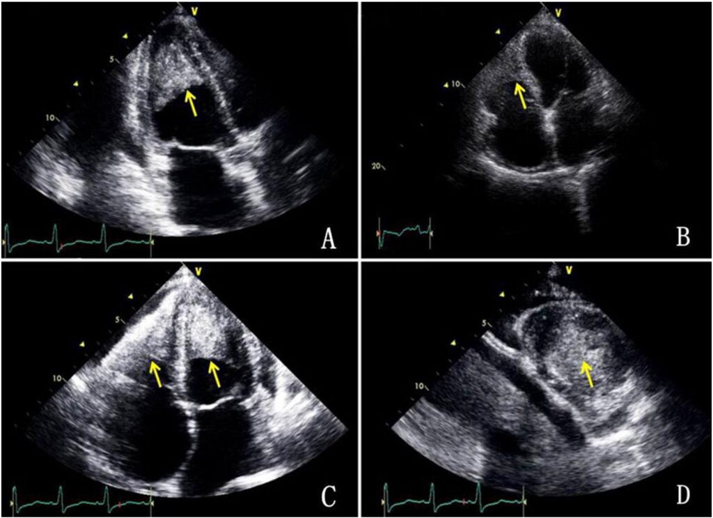
Benign masses were more frequently pedunculated, mobile, and adherent to the interatrial septum (p<0.001). Malignant masses showed a greater diameter and exhibited a higher frequency of irregular margins, an inhomogeneous appearance, sessile implantation, polylobate shape, and pericardial effusion (p<0.001). Infiltration, moderate-severe pericardial effusion, non-left localization, sessile, polylobate, and inhomogeneity were confirmed to be independent predictors of malignancy in both univariate and multivariable models. The predictive ability of the unweighted count of >3 was very high (>0.90) and similar to that of the previously published weighted score. The CTA generated an algorithm in which infiltration was the best discriminator of malignancy, followed by non-left localization and sessile shape. The percentage correctly classified by the CTA as malignant was 87.5%. Agreement between observer readings and cardiac mass histology ranged between 85.1-91.5%. The presence of at least 3 echocardiographic parameters was associated with a lower survival.
Conclusions
In the approach to CM, some echocardiographic parameters can serve as markers to accurately predict malignancy, thereby informing the need for second-level investigations and minimizing the diagnostic delay in such a complex clinical scenario.


